Digital dentistry
3D X-rays & Cone-Beam CT
Understand when a three-dimensional view can help treatment planning.
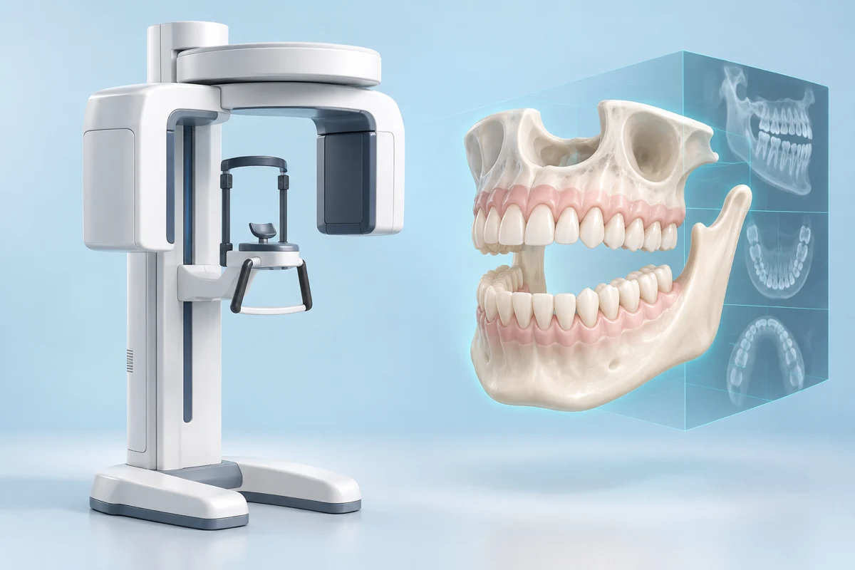
What it does
Cone-beam computed tomography (CBCT) uses X-rays to reconstruct a 3D view of teeth and jaws. It can help answer selected questions in implant planning, root canal care and oral surgery.
Explore the source material ↗Watch the explanation
See the procedure in motion
From positioning to a 3D dental X-ray
Planmeca
Connects to Vimeo when you choose to load.From positioning to a 3D dental X-ray
Watch a Viso CBCT scan being captured with Romexis software, including positioning and the imaging workflow.
Manufacturer training demonstration. The scan area and radiation settings must be selected for the patient’s clinical need.
If the player is unavailable, use the direct video link. Videos are external educational resources, not endorsements or personalized treatment advice. Publishers may include advertising; availability and captions vary. Loading connects to the video host and may share connection information or use cookies. Privacy notice.
What usually happens
The sequence and number of visits depend on the individual case.
Identify the question
The dentist decides whether existing records or lower-dose imaging can answer the clinical question.
Select the scan
The scan area and exposure are chosen for the information needed.
Capture the images
The unit rotates around the head while you remain still.
Interpret the result
A qualified clinician reviews the scan and explains findings relevant to your care.
Benefits, limits & trade-offs
CBCT generally exposes patients to more radiation than conventional dental X-rays. It should be justified by a clinical need, not offered automatically because an office has the equipment.
Watch & explore further
Open the publisher’s page for videos, animations or more detailed reading.
External media opens only when you choose a link. Publishers may show advertisements or use cookies. Links do not imply endorsement or a partnership. Illustrations on Teeth.com are AI-generated, simplified explanations, not patient images.
Questions for your dentist
- What question will this scan answer?
- Could existing or lower-dose images answer it?
- Who will interpret the entire scan and explain any unexpected findings?
Take the next step
Look for offices offering this care
Search for 3d x-rays & cone-beam ct in your area, then confirm the service with the office.
Opening Google Maps shares your search terms with Google. Results and rankings come from Google. Teeth.com has not verified these offices, their equipment, training or availability. A listing is not a recommendation.
Before you book
- Confirm that the office offers the specific procedure or technology you want to discuss.
- Ask who performs the treatment and about their relevant training and experience.
- Discuss whether it suits your situation, alternatives, the total fee and follow-up care.